January 25, 2023

Pectus excavatum: an old problem
As many as 1 in 400 people have pectus excavatum, a depression deformity of the chest. Some children present early in life while many will first notice deepening of the sternum around the onset of puberty. Commonly, those affected have not been offered work up or treatment; female patients are especially under diagnosed, as their breast tissue can make it difficult to assess the severity of the deformity. Through social media, many teenagers and young adults are finding information – good and bad – on chest wall deformities online. One man on TikTok has a video of his goldfish swimming around his severely depressed chest; others have published videos of workouts intended to ‘correct’ pectus excavatum. Devices are sold that claim to elevate the sternum. With our patients self-diagnosing this condition, it is worth reviewing the current evidence including work-up and treatment options.
Physiology and symptoms
The depression of the sternum has physiological consequences. For years, compression of the lungs was thought to be a primary reason for symptoms. However, intra-operative echocardiogram studies have shown that the sternum compresses the right heart, leading to decreased stroke volume, and valvular abnormalities. This manifests primarily as exercise intolerance. Many patients believe they have exercise induced asthma, when in reality, their shortness of breath during physical activity is due to their inability to maintain adequate cardiac response, especially with prolonged activity. Patients seen at our center will commonly report an inability to keep up with their peers, despite being in otherwise good physical health. They may also report chest pain, palpitations, and less commonly, dysphagia. Unfortunately, some patients also report being teased or bullied due to their condition.
Surgical treatment is the gold standard for severe pectus excavatum. However, there is a historical bias against surgical repair – perhaps rightly so, as it took many years for surgeons to come up with a safe, durable surgery, and, to decide who would benefit from repair.
A brief history
Hippocrates first described deformities of the chest wall. No treatments were undertaken until the 20th century. With the advent of anesthetic agents, the field of surgery expanded the number and types of conditions it treated. Early surgical attempts at correction of pectus excavatum in children included costal cartilage resection and placing the chest in traction, seeking to elevate the depressed sternum. Then in 1949, Dr. Mark Ravitch introduced a method of repair which expanded on previous efforts, fully mobilizing the sternum from all attachments. This procedure, or variations of it, became standard of care. However, many young children underwent this correction, and went on to develop asphyxiating chondrodystrophy. Watching children suffer from complications of the surgery led to push back from pediatricians – the treatment of pectus excavatum was labelled worse than the disease. Thus, children and their parents were commonly told that the condition had no good treatments, or, that pectus excavatum is a cosmetic condition.
New(-er) evidence
In 1986, Dr. Donald Nuss, a pediatric surgeon at Children’s Hospital of the King’s Daughters in Virginia, realized that the sternum could be elevated without removing existing tissue. He took advantage of the flexibility of the chest, and inserted a titanium bar under the sternum, fully correcting the excavatum. Since that first repair, thoracoscopy has been added, as well as other modifications to increase the safety and durability of the Nuss procedure, also know as Minimally Invasive Repair of Pectus Excavatum (MIRPE). The Nuss procedure is now internationally accepted as the standard of care for correction of severe pectus excavatum.
What is severe? The work up
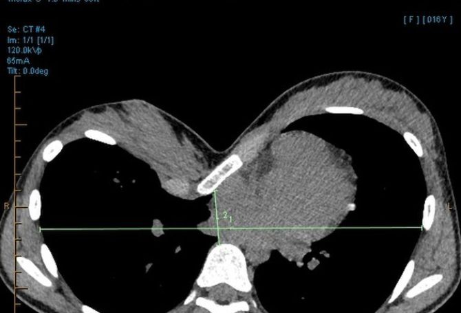
When evaluating patients with this deformity, in addition to a detailed history and examination of their chest, we examine for evidence of associated problems such as scoliosis, Poland syndrome, and connective tissue disorders including Marfan’s and Ehlers-Danlos. In teenagers, additional work up quantifies the severity during the decision making for surgery. Echocardiogram assesses for cardiac compression and other abnormalities. Cross sectional imaging such as MRI / CT will quantify the Haller Index: the internal transverse diameter of the chest divided by the anterior-posterior measurement from the back of the sternum to the front of a vertebral body. A Haller Index of 3.25 or higher defines a severe case of pectus excavatum.
Modern, safe treatment
Patients with severe pectus excavatum are candidates for the Nuss procedure or MIRPE. Surgery is often done in the early teen years, though can also be offered to adult patients who were not treated in the teen years. One or more metal bars are placed into the chest, elevating the sternum, and relieving the cardiac compression. Multi-modal pain relief, including cryoablation of the intercostal nerves, has greatly improved surgical recovery, with a one-night average length of stay.
This hardware usually stays in place for three years. The average patient misses about two to three weeks of school, is restricted from contact sports for three months after surgery and has a durable long-term result after surgery.
When to refer
Many surgeons would like to see patients with pectus deformities (excavatum and carinatum) beginning around age 10, or, at disease onset. We will follow annually, to assess whether the sternum becomes deeper with puberty and to follow symptoms. Imaging is generally not done at diagnosis, rather, it is done when the patient is nearly ready for surgery. If cross sectional imaging is done too early, it can underrepresent the severity of the defect, which commonly worsens during puberty.
In summary, there is a safe and effective treatment for teens and adults with pectus excavatum. Failure to treat can lead to long-standing exercise intolerance. Feel free to reach out with questions – our program seeks to eliminate barriers to care for these patients.
Kim Ruscher, MD MPH
kruscher@peacehealth.org